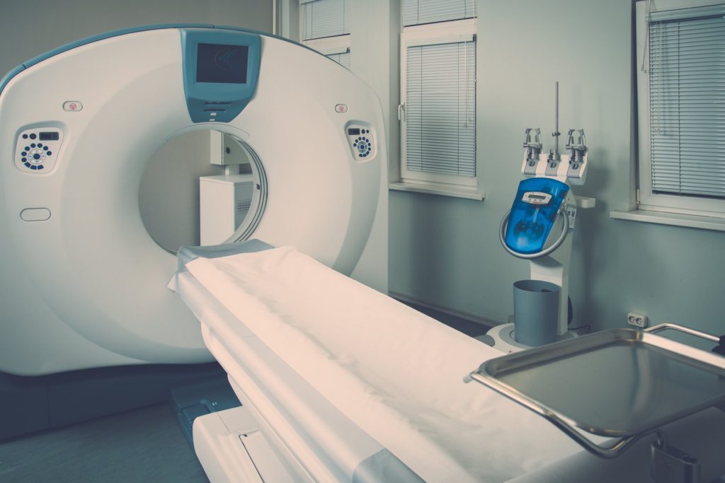
PET/CT scanners are a combination of two of the most distinctive medical imaging systems. PET/CT scanners have become one of the most prominent and active imaging procedures known to-date. PET/CT can be defined as an advanced nuclear medical imaging technique. The PET/CT technique uses a combination of molecular imaging known as Positron Emission Tomography (PET) and Computed Axial Tomography (CT). The combination of both technologies is then used for one of the most sophisticated and thorough medical imaging processes.
PET scans are advanced imaging systems that are designed for revealing a great deal of information regarding the function of cells and tissues in a patient’s body. PET itself offers significant detail. However, when PET is combined with CT, it allows the ability to focus on the structure of the internal anatomy. A PET/CT scan can then, collect and produce an unparalleled amount of data in a single scan.
The PET nuclear imaging technique provides the visualization of processes that are metabolic in the body. These scans are minimally invasive and are generally painless to patients. A PET scan uses radioactive materials known as radiopharmaceuticals. These materials can be injected into a patient’s vein, inhaled as a gas, or even ingested orally. The radiopharmaceuticals (also referred to as tracers), is part of a glucose solution which can be absorbed and processed by organs or tissues in the patient’s body when being examined. When inside the body, these tracer particles emit rays that are then finely tuned by the PET systems detector panels which can track and distinguish the inside of the body.
For the part of the body that is being observed, the system’s software would be able to render the bodily function clearly for diagnosis and observation. By combining the PET data with Computed Tomography, the system would then be able to develop 3-D images of the concentration provided by the tracer and their interactions with tissues surrounding the area. The data from the two exams can be correlated and combined into a single image which leads to more precise information for medical experts.
While there may be a difference in the cost of PET/CT scanners depending on the location, the scanning experience is quite similar.
Generally, radiologists would use PET/CT scans for locating and determining cancerous tissue in patients. This is because cancerous cells are known to use more glucose than regular cells and doctors would be able to identify the grade of the tumor by examining the rate using tracer infused glucose solution. Additionally, PET/CT scanners would also be able to help doctors determine how effective chemotherapy treatments would be for cancer patients.
Other major uses of PET/CT scans include:
• The system can track the blood flow to the heart and other muscles.
• The effects of a heart attack on patients.
• The areas of the heart that can benefit from heart surgery.
If you’re looking obtain more information on a PET/CT scanner purchase for your medical facility, we at Amber USA are more than happy to help. We issue a variety of used and refurbished medical imaging equipment including multiple brands of PET/CT scanners plus many other second hand medical equipment (Philips CT scanner, Siemens MRI machines), offering you a wide range of options to choose from.
If you were looking for a reliable place that offers used medical equipment for sale for budget prices, you have come to the right place. Contact us now!